
Medically reviewed by C.H. Weaver M.D. Medical Editor updated 8/2025
- Introduction to Gastric Cancer
- Symptoms & Signs
- Causes & Risk Factors
- Diagnosis & Tests
- Stages of Gastric Cancer
- Prevention & Screening
Introduction to Gastric Cancer
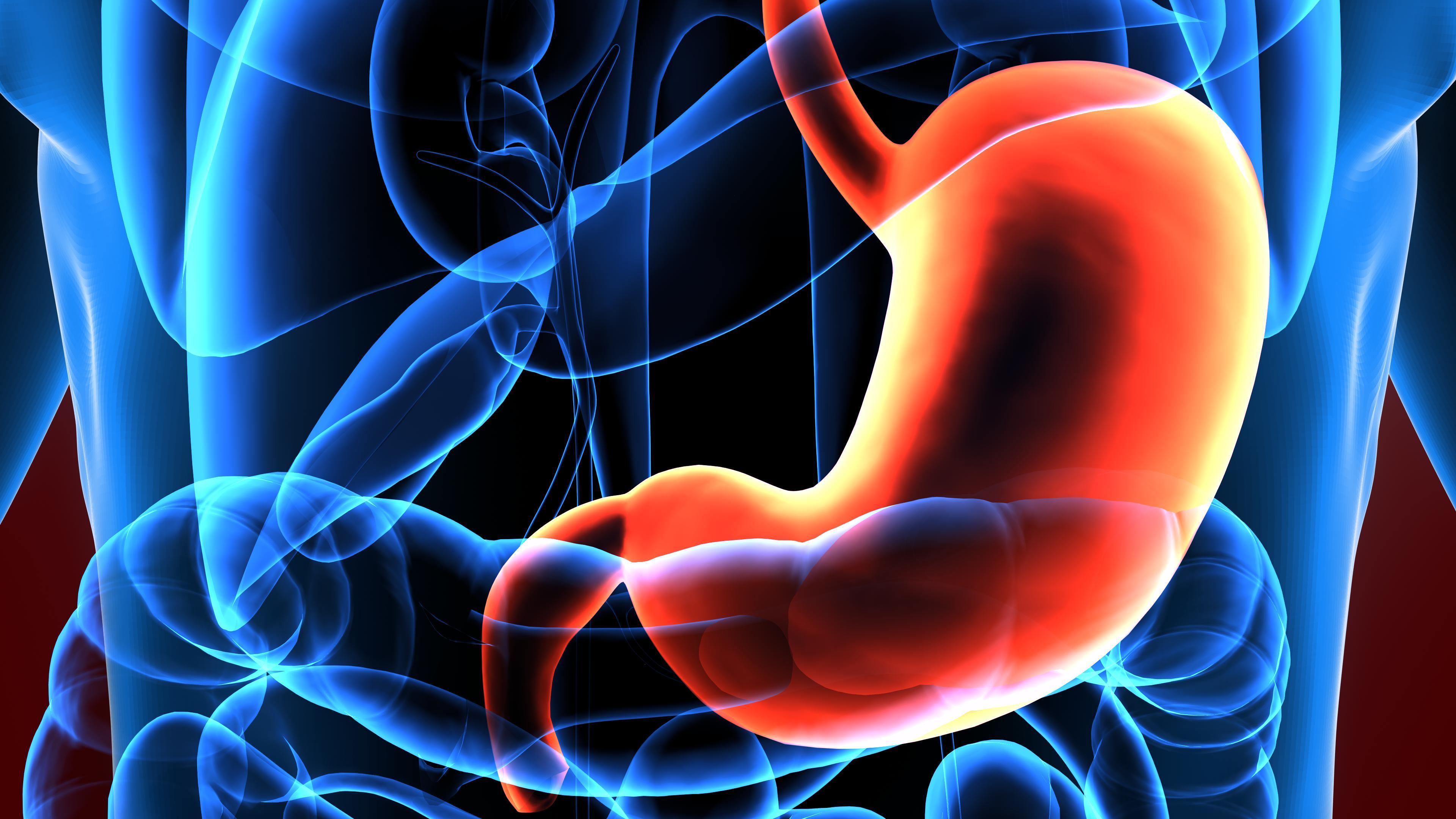
The stomach is the part of the digestive system responsible for digesting food. Food passes through the esophagus into the stomach at the level of the diaphragm, the breathing muscle that separates the abdomen from the chest. The stomach extends from the diaphragm to the first portion of the small intestine.
Cancer of the stomach is called gastric cancer. Gastric adenocarcinoma is the most common type of gastric cancer and arises from the cells lining the surface of the stomach.
Over the past two to three decades, incidences of gastric cancer have significantly declined in the United States and many other industrialized countries. In the U.S., stomach cancer now represents approximately 1.5% of all newly diagnosed cancers each year.4 An estimated 30,300 individuals are diagnosed annually, with a higher prevalence in men (17,720 cases) compared to women (12,580 cases). Each year, gastric cancer results in approximately 10,780 deaths nationwide.4
Symptoms & Signs
Individuals with gastric cancer may experience the following symptoms or signs.10 Sometimes, people with gastric cancer do not have any of these changes, or the cause of a symptom may be another medical condition besides cancer.
- Bloating
- Indigestion or heartburn
- Vomiting
- Nausea
- Pain or discomfort in the abdomen
- Diarrhea or constipation
- Bloating of the stomach after meals
- Loss of appetite
- Sensation of food getting stuck in the throat while eating
Symptoms of advanced stomach cancer may include:
- Weakness and fatigue
- Vomiting blood or having blood in the stool
- Unexplained weight loss
Causes & Risk Factors
Gastric cancer begins when healthy cells in the stomach acquire a genetic change (mutation) that causes them to turn into abnormal cells capable of rapidly dividing.
A risk factor is anything capable of increasing an individual’s likelihood of developing cancer from cellular mutations. While risk factors can influence cancer development, they do not typically cause cancer directly. Moreover, not everyone with one or more risk factors will develop cancer, and some individuals with no known risk factors may still be diagnosed with the disease.
Most gastric cancers arise sporadically, without a known cause. However, some cases are more likely to occur in individuals with certain risk factors that increase the likelihood of developing the disease.
The primary risk factor associated with gastric cancer is chronic inflammation due to infection with bacterium Helicobacter pylori (H. pylori). H. pylori infection is common, but most people who are infected do not develop gastric cancer. The bacterium may be spread from person to person as well as through contact with contaminated food or water. Infection with H. pylori causes a reduction in the normal acid production and a shrinkage and loss of cells (called atrophy) in the stomach. It can take decades for the precancerous conditions caused by H. pylori infection to develop into cancer. In addition, H. pylori can be treated with antibiotics, which may reduce the risk of gastric cancer.3
However, a number of other factors may raise a person’s risk for developing gastric cancer.5
Genetic Factors
The chance of an individual developing cancer depends on both genetic and non-genetic factors. A genetic factor is an inherited, unchangeable trait. Someone with a genetic predisposition may be at higher risk for a certain cancer if a family member has that type of cancer. Gastric cancer can sometimes be linked to inherited genetic factors, with approximately 1%–3% of cases arising from hereditary syndromes.. These conditions increase lifetime cancer risk and often require specialized surveillance or preventive measures. Table 1 outlines syndromes, associated genes and gastric cancer risk.Table 1: Major Hereditary Syndromes Linked to Gastric Cancer11
| Hereditary Syndrome | Gene | Gastric Cancer Risk |
| Hereditary diffuse gastric cancer4,6,8,9 | CDH1 and CTNNA1 | 33%–83% |
| Familial adenomatous polyposis syndrome (FAP)1,7 | APC | 1.3%–5% in FAP and AFAP |
| Attenuated familial adenomatous polyposis (AFAP) | APC | |
| Gastric adenocarcinoma and proximal polyposis of the stomach (GAPPS) | APC (pathogenic variants/single nucleotide variant in the promoter 1B region of APC are associated with GAPPS-7) | In GAPPS, gastric cancer risk is significantly increased, but exact risk numbers are unknown |
| Lynch syndrome (also known as hereditary nonpolyposis colorectal cancer [HNPCC]) | MLH1, MSH2, MSH6, PMS2, EPCAM | 0.2%–9% |
| Peutz-Jeghers syndrome | STK11 (also known as LKB1) | 29% |
| Juvenile polyposis syndrome | SMAD4, BMPR1A | 21% if multiple polyps are present |
| Li-Fraumeni syndrome | TP53 | 2.8% |
Emerging Genetic Links: Research suggests potential roles for homologous recombination genes (BRCA1, BRCA2, PALB2) in gastric cancer risk and other genes are currently being studied.
Non-Genetic Environmental Factors
A non-genetic factor is a variable in a person’s environment, which can often be changed. Some non-genetic factors play a role in facilitating the process of healthy cells turning cancerous (e.g. the correlation between smoking and lung cancer). Other cancers have no known environmental correlation but are known to have a genetic predisposition. Common examples include:
- Using any form of tobacco: cigarettes, cigars, pipes, chewing tobacco, and snuff increases the risk of gastric cancer.
- Heavy drinking over a long period of time increases the risk of developing gastric cancer
- Being severely overweight and having too much body fat can increase a person’s risk.
- Eating a diet high in salt has been linked to an increased risk of stomach cancer. This includes foods preserved by drying, smoking, salting, or pickling and foods high in added salt.
- Occupational exposure. Exposure to certain dusts and fumes may increase the risk of developing stomach cancer.
Additional Risk Factors
- Stomach cancer occurs most commonly in people older than 55. Most people diagnosed with stomach cancer are in their 60s and 70s.
- Men are twice as likely to develop stomach cancer as women.
- Race/ethnicity. Stomach cancer is more common in black, Hispanic, and Asian people than in white people.
- Previous surgery or health conditions. People who have had stomach surgery, pernicious anemia, or achlorhydria have a higher risk of stomach cancer.
- Atrophic Gastritis.
Diagnosis & Tests
Doctors can use many tests to find and diagnose cancer. They can also perform tests to learn if cancer has spread from where it originated.
A biopsy remains the only definitive method for diagnosing gastric cancer. During this procedure, a doctor removes a small sample of stomach tissue, usually through upper endoscopy, for laboratory analysis.
While biopsy confirms the presence of cancer, accurately determining the stage and extent of the cancer is more complex. Current clinical staging methods are imperfect and continue to evolve with advances in diagnostic technology. In many cases, the extent of cancer spread is best assessed after surgical evaluation, as preoperative tests may not fully capture the disease’s progression.
To help evaluate and stage gastric cancer, a variety of diagnostic procedures may be used. Listed below are some of the more common techniques for assessing gastric cancer:
- Upper endoscopy or gastroscopy (EGD): An EGD is the most commonly used technique used to diagnose gastric cancer. It is an examination performed with an endoscope, a thin, flexible tube with a light and camera. The endoscope is inserted through the mouth into the esophagus and allows the physician to visualize, photograph, and biopsy the suspicious areas for cancer. It allows the physician to see abnormal areas and take biopsies.
- Computed Tomography (CT) Scan: A CT scan is a technique for imaging body tissues and organs, during which X-ray transmissions are converted to detailed images, using a computer to synthesize X-ray data. A CT scan is conducted with a large machine positioned outside the body that can rotate to capture detailed images of the organs and tissues inside the body.
- Endosonography: Endosonography refers to an ultrasound test performed through an endoscope. Ultrasound tests utilize sound waves to detect different densities of tissue, including cancer. Endosonography can detect spread of cancer into various layers of the stomach, adjacent organs and lymph nodes better than CT scanning.
- Laparoscopy: Laparoscopy is a procedure that involves the insertion of an endoscope through a small incision in the abdomen. Laparoscopy is an important tool for staging and has proven to be more reliable than CT scanning in detecting spread of cancer to the liver and the lining of the abdomen (peritoneum).
- Magnetic Resonance Imaging (MRI): MRI uses a magnetic field rather than X-rays, and can often distinguish more accurately between healthy and diseased tissue than a CT. An MRI gives a better picture of cancer located near bone than does CT, does not use radiation, and provides pictures from various angles that enable doctors to construct a three-dimensional image of the cancer.
- Positron emission tomography (PET): Positron emission tomography scanning is an advanced technique for imaging body tissues and organs. One characteristic of living tissue is the metabolism of sugar. Prior to a PET scan, a substance containing a type of sugar attached to a radioactive isotope (a molecule that emits radiation) is injected into the patient’s vein. The cancer cells “take up” the sugar and attached isotope, which emits positively charged, low energy radiation (positrons) that create the production of gamma rays that can be detected by the PET machine to produce a picture. If no gamma rays are detected in the scanned area, it is unlikely that the mass in question contains living cancer cells.
Biomarker Testing: A biomarker is a gene, protein, or other substance found in blood, body fluids or tissue that can help with detection and understanding of a tumor. Cancer biomarkers can be produced by the cancer itself or by the body in response to cancer. Biomarkers are used in several ways:
-
- Diagnosis: To help detect or confirm the presence of cancer.
- Prognosis: To predict how aggressive the cancer might be or how it may behave over time.
- Treatment Guidance: To help determine the most effective, personalized treatments such as targeted therapies or immunotherapies.
- Monitoring: To track how well treatment is working or if the cancer has returned.
Relevant markers in gastric cancer include microsatellite instability (MSI) or mismatch repair (MMR) proteins, HER2 overexpression/amplification, PD-L1 expression, and gene alterations like RET, NTRK, or BRAF. Testing for these biomarkers can help guide treatment decisions by determining whether targeted therapies or immunotherapies may be effective. Universal testing for MSI or mismatch repair (MMR) should be performed for all newly diagnosed gastric cancers.
If a cancer is unresectable, recurrent, or metastatic, HER2 or PD-L1 expression testing as well as tumor mutation testing are recommended. Testing is done using a variety of methods such as fluorescence in situ hybridization (FISH), immunohistochemistry (IHC), next-generation sequencing (NGS), and/or polymerase chain reaction (PCR). Learn more about biomarker testing here.
Repeat Biomarker Testing: In patients whose cancer progresses after treatment, repeat biomarker testing may be warranted to determine if changes in the tumor biology could impact treatment selection.
Gastric Cancer Staging
In addition to biomarker testing, it is important to determine the stage of the cancer, or how much it has spread, before initiating treatment. Staging describes:
- Tumor size and location
- If the tumor has grown through the layers of the stomach wall, and
- If cancer has spread to lymph nodes, organs, or other parts of the body.
The stage of cancer plays a critical role in guiding treatment decisions, as advanced stages with widespread disease may make complete surgical removal impossible.
Unfortunately, in many cases, the true extent of spread of gastric cancer can only be determined at the time of surgical resection. Frequently, more advanced cancer is found during surgery than was detected through diagnostic procedures.
In order to learn more about the most recent information available concerning the treatment of gastric cancer, select from the following.
Early-stage gastric cancer
Loco-regional gastric cancer
Metastatic or recurrent gastric cancer
For more information about how gastric cancer is treated, read here.
Prevention & Screening
Prevention of Gastric Cancer
About two-thirds of cancer deaths in the U.S. are linked to tobacco use, poor diet, obesity, and lack of physical activity. Understanding how risk factors may increase the odds of getting cancer is a crucial step toward prevention. However, the potential to reduce cancer risk through lifestyle changes remains underrecognized. Some of the most effective ways to help prevent gastric cancer include:
- Treatment of H. pylori infection. According to studies conducted in parts of the world where gastric cancer is common, treatment of H. pylori infections may reduce the risk of gastric cancer. In a large combined analysis of people infected with H. pylori, gastric cancer developed in 1.1% of those who received treatment to eradicate the infection and 1.7% of those who did not receive treatment.3
- Aspirin is a type of non-steroid anti-inflammatory drug (NSAID). Studies have suggested that regular use of aspirin—but not other types of NSAIDs—may reduce the risk of gastric cancer.2 Because regular aspirin use also carries some risks, people who are considering taking aspirin on a regular basis are advised to talk with their physician.
- Diet is a fertile area for immediate individual and societal intervention to decrease the risk of developing certain cancers.
- Avoidance of tobacco smoke. Smoking increases the risk of many types of cancer, including gastric cancer. Never smoking (or quitting if you’ve started) provides many health benefits.
Screening and Early Detection of Gastric Cancer
Gastric cancer rates have been steadily declining in the United States and at this point, routine screening for this cancer is not performed in individuals without significant risk factors. For individuals who have a family history of gastric or related cancers, it is important to consult with a genetic counselor to assess testing eligibility and personalized risk management. Early detection and preventive strategies significantly improve outcomes for high-risk individuals.
Mass screening programs for gastric cancer have been effective in high-risk areas, such as Japan. In some Japanese studies, as many as 40% of newly diagnosed patients have early gastric cancer and as many as 60% of patients are actively participating in mass screening programs. Routine screening in Japan involves gastroscopy.
References:
- Cannon AR, Keener M, Neklason D, et al.: Surgical Interventions, Malignancies, and Causes of Death in a FAP Patient Registry. J Gastrointest Surg 25 (2): 452-456, 2021.
- Epplein M, Nomura AMY, Wilkens LR, Henderson BE, Kolonel LN. Nonsteroidal anti-inflammatory drugs and risk of gastric adenocarcinoma: The Multiethnic Cohort Study. American Journal of Epidemiology. 2009; 170:507-14.
- Fuccio L, Zagari RM, Eusebi LH et al. Meta-analysis: can Helicobacter pylori eradication treatment reduce the risk for gastric cancer? Annals of Internal Medicine. 2009;151:121-8.
- Hansford S, Kaurah P, Li-Chang H, et al.: Hereditary Diffuse Gastric Cancer Syndrome: CDH1 Mutations and Beyond. JAMA Oncol 1 (1): 23-32, 2015.
- Lavin TP, Weitzel JN, Neuhausen SL, Schrader KA, Oliveira C, Karam R. Genetics of gastric cancer: what do we know about the genetic risks? Trans Gastroenterol Hepatol. 2019 Jul 29;4:55.
- Kaurah P, MacMillan A, Boyd N, et al.: Founder and recurrent CDH1 mutations in families with hereditary diffuse gastric cancer. JAMA 297 (21): 2360-72, 2007.
- Mankaney G, Leone P, Cruise M, et al.: Gastric cancer in FAP: a concerning rise in incidence. Fam Cancer 16 (3): 371-376, 2017.
- Pharoah PD, Guilford P, Caldas C, et al.: Incidence of gastric cancer and breast cancer in CDH1 (E-cadherin) mutation carriers from hereditary diffuse gastric cancer families. Gastroenterology 121 (6): 1348-53, 2001.
- Roberts ME, Ranola JMO, Marshall ML, et al.: Comparison of CDH1 Penetrance Estimates in Clinically Ascertained Families vs Families Ascertained for Multiple Gastric Cancers. JAMA Oncol 5 (9): 1325-1331, 2019.
- Signs and Symptoms of Stomach Cancer | Gastric Cancer Warning Signs. (2024, October 2). American Cancer Society. Retrieved June 9, 2025, from https://www.cancer.org/cancer/types/stomach-cancer/detection-diagnosis-staging/signs-symptoms.html
- Stomach (Gastric) Cancer Key Statistics. (2025, January 16). American Cancer Society. Retrieved June 4, 2025, from https://www.cancer.org/cancer/types/stomach-cancer/about/key-statistics.html
- What Are the Risk Factors for Stomach Cancer? (2021, January 22). American Cancer Society. Retrieved June 4, 2025, from https://www.cancer.org/cancer/types/stomach-cancer/causes-risks-prevention/risk-factors.html
- Xicola RM, Li S, Rodriguez N, et al.: Clinical features and cancer risk in families with pathogenic CDH1 variants irrespective of clinical criteria. J Med Genet 56 (12): 838-843, 2019.


