
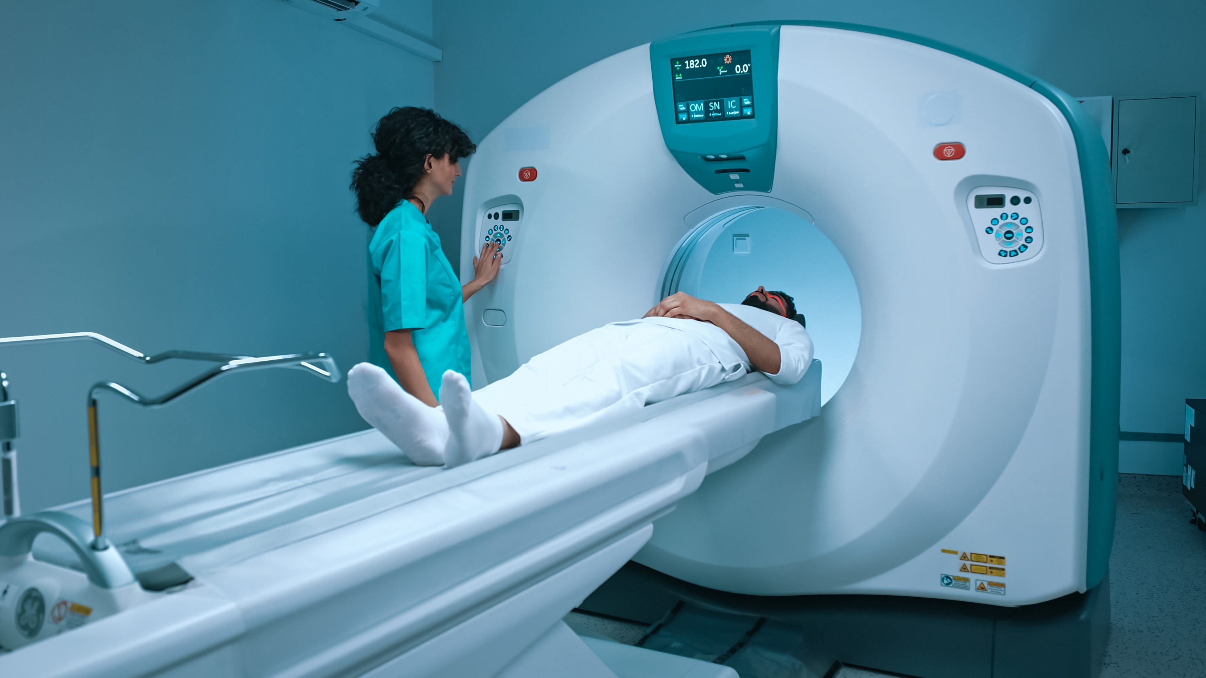
A recent retrospective cross-sectional study has found that many cases of high-risk prostate cancer may be more advanced than previously thought. The study used an advanced imaging technique called Prostate-Specific Membrane Antigen (PSMA)-PET to detect cancer spread in patients previously classified as having non-metastatic disease.
Study Background
A total of 182 patients with high-risk non-metastatic hormone-sensitive prostate cancer who met inclusion criteria of the EMBARK clinical trial were analyzed from 4 prospective study database that included 2002 patients. The treatment history of the 182 patients with prostate cancer that was thought to be confined to the prostate (non-metastatic) and sensitive to hormone therapy included:
- Half of the patients (91) had undergone surgery to remove their prostate (radical prostatectomy).
- About one-fifth (39) had received definitive radiation therapy as their primary treatment.
- The remaining 29% (52) had received salvage radiation therapy after their prostate surgery.
Key Findings
- Nearly half of high-risk prostate cancer patients thought to have localized disease actually had metastases when examined with PSMA-PET imaging
- 24% of patients showed 5 or more lesions that were missed by conventional imaging
- PSMA-PET detected cancer metastases in 46% of patients previously thought to have no cancer spread
PSMA-PET imaging uses radioactive tracers that bind to prostate cancer cells, making them visible on PET scans. This allows doctors to see the biological activity of the cancer, not just anatomical details. The investigators from the study emphasized that PSMA-PET plays a critical role in accurately staging prostate cancer, which can significantly impact treatment decisions and outcomes. The findings challenge the results of previous clinical trials that relied on conventional imaging, such as the EMBARK trial. Researchers suggest that future trials should include PSMA-PET for patient selection to ensure more accurate results.
While more research is needed to fully understand the impact of PSMA-PET on long-term patient outcomes, the researchers who conducted this study are confident that this advanced imaging technique will continue to improve prostate cancer staging and guide personalized therapies.
About PSMA-PET
The imaging agent used in PSMA-PET imaging was approved by the FDA in December, 2021. but currently has limited availability in the US.
A PSMA PET scan is an advanced imaging technique currently used in three main scenarios for prostate cancer patients:
- Checking for Cancer Spread in Potentially Curable Cases
- Used to rule out metastases (cancer spread) beyond the pelvis
- Helps determine if surgery or radiotherapy are suitable options
- Identifies patients who may need whole-body treatments instead
- Detecting Cancer Recurrence
- For patients who’ve had surgery or radiotherapy but show rising PSA levels
- Helps guide decisions between additional radiotherapy or systemic therapy
- Assessing Eligibility for Lu-177 PSMA Treatment
- Used for patients whose cancer has progressed despite chemotherapy and hormone treatments
- Determines if the cancer expresses enough PSMA for this targeted therapy to be effective
- Helps avoid ineffective treatments in patients with low PSMA expression
This imaging technique plays a crucial role in personalizing treatment plans and ensuring patients receive the most appropriate care for their specific situation. Patients with prostate cancer can talk to their doctor about the risks and benefits of a PSMA-PET.
More Reading
Treatment & Management of Prostate Cancer
Join the Conversation on CancerConnect!
Reference
Holzgreve A, Armstrong WR, Clark KJ, et al. PSMA-PET/CT Findings in Patients With High-Risk Biochemically Recurrent Prostate Cancer With No Metastatic Disease by Conventional Imaging. JAMA Netw Open. 2025;8(1):e2452971.
Kwon DH, Velazquez AI, de Kouchkovsky I. PSMA PET Scan. JAMA Oncol. 2022;8(12):1860.


