
by Dr. C.H. Weaver 3/2023
On March 9, 2023 the U.S. Food and Drug Administration published updates to mammography regulations requiring mammography facilities to notify patients about the density of their breasts. The final rule amends regulations issued under the Mammography Quality Standards Act (MQSA) of 1992, a law passed to ensure quality mammography, which is very important for early breast cancer detection. The MQSA authorizes FDA oversight over mammography facilities, including their accreditation, certification, annual inspections to help ensure mammography facilities provide quality care. One of the key updates to regulations under the MQSA requires facilities to provide information to patients regarding the density of their breasts.

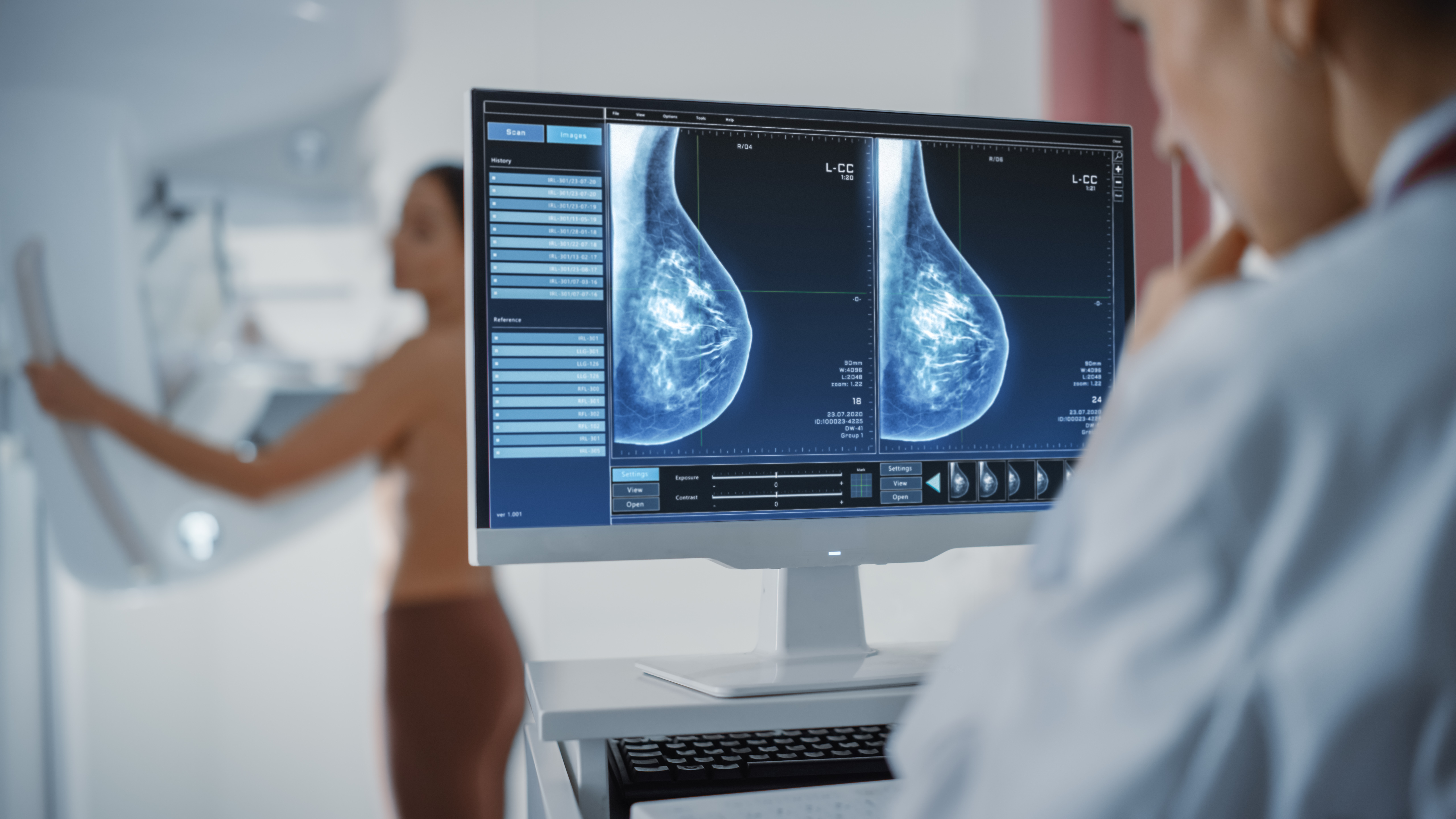
What is breast density?
Breast density refers to the tissue composition of a breast. All breasts contain a mixture of fatty and glandular (dense) tissue. The more glandular tissue present, the “denser” the breast is considered. Dense breasts are normal and occur in ~ 40% of women of mammography age. The denser the breast, the greater the risk of breast cancer and a “normal” or “negative” mammogram may not always reliably exclude a cancer.
Why does breast density matter?
Breast density is important because it is associated with an increased risk of developing breast cancer and to have that cancer missed by mammography.1,2
This is because both dense breast tissue and breast cancer display as white or grey on a mammogram which makes finding a cancer in a dense breast more difficult. Cancer in a fatty breast is easier to detect as the fatty tissue displays darker and the cancer appears white. In a dense breast, little or no contrast may be present. Thus, as breast density increases, a mammogram’s ability to show cancer decreases.
Is breast density associated with worse outcomes?
High breast density on mammogram is associated with an increased risk of developing cancer, bit it does not appear to increase the risk of death among breast cancer patients,3 according to a study evaluating over 9000 women with breast cancer. In fact the researchers found an increased risk of breast cancer death associated with lower breast density—especially among obese patients.
Role of Magnetic Resonance Imaging
Magnetic resonance imaging (MRI) is the best supplemental imaging modality for detecting breast cancer in women with dense breasts and negative mammography, according to a meta-analysis of 22 studies encompassing 261,233 patients, 120,081 of whom had dense breasts and a negative mammogram.5
Breast MRI was found to be superior for incremental cancer detection compared with handheld breast ultrasound, automated whole-breast ultrasound, and digital breast tomosynthesis.
According to the study authors the pooled data showed that MRI was the best supplemental imaging modality in women at average risk or intermediate risk for breast cancer with dense breasts and mammography negative for cancer.
Annual Mammograms Important
Although it is recommended that women between the ages of 40-49 with dense breasts require annual screening mammography to increase the rate of detection of breast cancer the optimal interval for screening mammography continues to be evaluated. Some researchers believe that younger women may benefit from more frequent screening.
Researchers from the University of Washington evaluated 576 women who were diagnosed in between mammography screenings (interval cancers), with intervals of either 12 or 24 months between screenings. When mammography was performed every 12 months, a diagnosis of an “interval cancer” was found to occur in almost 30% of younger women, compared to only 14% of older women. When mammography was performed every 24 months, diagnosis of interval cancers occurred in over 50% of younger women, and 25% of older women. The researchers concluded that annual screening mammography in women 40-49 years increases the rate of cancer detection compared to every 2 years, mainly due to breast density and rapid cancer growth. Women who are at a high risk of developing breast cancer may need to be on a more frequent screening schedule. Patients should speak with their physician about their individual risks and benefits of specific screening schedules for the early detection of breast cancer.

Women with dense breast should discuss the role of annual mammography especially if they are 40-49 years old.4 and whether MRI in addition to mammography should be considered.
References:
- Barlow WE, White E, Ballard-Barbash R et al. Prospective Breast Cancer Risk Prediction Model for Women Undergoing Screening Mammography. Journal of the National Cancer Institute. 2006;98:1204-14.
- Chen J, Pee D, Ayyagari R et al. Projecting Absolute Invasive Breast Cancer Risk in White Women with a Model that Includes Mammographic Density. Journal of the National Cancer Institute. 2006;98:1215-26.
- Gierach GL, Ichikawa L, Kerlikowske K, et al. Relationship between mammographic density and breast cancer death in the breast cancer surveillance consortium. Journal of the National Cancer Institute. 2012; 104 (16): 1218-1227.
- Buist DSM, Porter PL, Lehman C, et al. Factors contributing to mammography failure in women aged 40-49 years. Journal of the National Cancer Institute. 2004;96:1432-1440.
- Hussein H, Abbas E, Keshavarzi S, et al. Supplemental breast cancer screening in women with dense breasts and negative mammography: A systematic review and meta-analysis. Radiology. Published online January 31, 2023. doi:10.1148/radiol.221785


