
What is FDG-PET Imaging?
Positron emission tomography (PET) is an imaging test used to show how your organs and tissues are working. Whereas other imaging tests, such as X-ray, CT, and MRI, reveal structural changes in the body, PET is used to reveal chemical and physiological changes. Uses for PET scans include checking brain function; diagnosing cancer, heart problems, and brain disorders; examining blood flow to the heart; and determining spread of cancer and response to therapy.
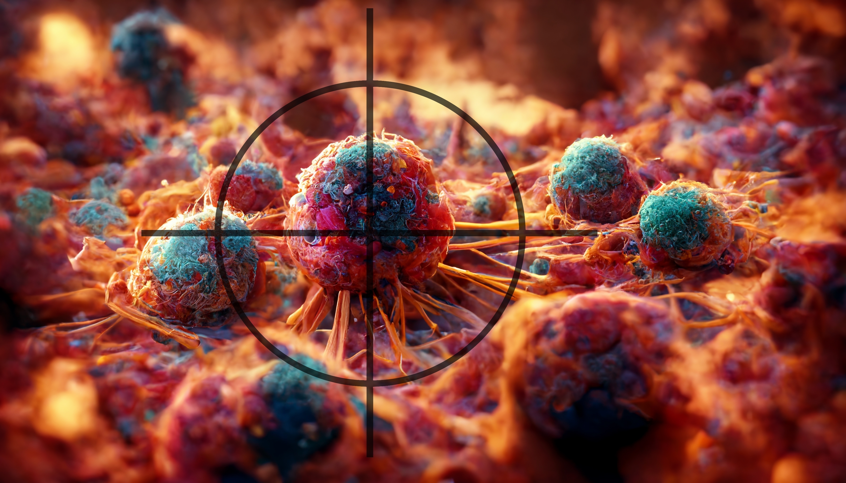
The use of PET scans may help doctors more accurately detect the presence and location of cancer cells. A PET scan is similar to a CT scan; however, PET scans can detect live cancer tissue.
Prior to a PET scan, the patient receives an injection of a substance that contains a type of sugar attached to a radioactive isotope (fluorodeoxyglucose positron emission tomography (FDG-PET)). The cancer cells “take up” the sugar and attached isotope, which emits positively charged, low energy radiation (positrons). The positrons react with electrons in the cancer cells, which create the production of gamma rays. The gamma rays are then detected by the PET machine, which transforms the information into a picture. If no gamma rays are detected in the scanned area, it is unlikely that the mass in question contains living cancer cells.
Understanding the Role of FES PET Imaging in ER+ Breast Cancer and Invasive Lobular Carcinoma
Patients with estrogen receptor-positive (ER+) breast cancer and/or invasive lobular breast cancer (ILC) may benefit from a newer imaging technique known as FES PET imaging. ILC can be challenging to detect with conventional imaging methods and FDG PET may underperform in the detection of ER+ lesions compared to FES PET.
What is FES PET Imaging?
18F-Fluorestradiol (18F-FES; also known by the brand name Cerianna) is an imaging agent approved by the FDA that helps assess the ER status of breast cancer lesions. It is used alongside PET imaging to identify ER-positive cancer areas in the body. Standard PET imaging alone is thought to underperform in low-grade ER-positive breast cancer. FES PET imaging involves the injection of a small amount of a radioactive substance that attaches to estrogen receptors in the cancer cells. This allows the radiologist to see where the cancer is and how active it is.
Why is FES PET Imaging Recommended?
According to the National Comprehensive Cancer Network (NCCN) Guidelines, FES PET imaging is recommended for evaluating estrogen receptor status during the staging process for patients with recurring or metastatic breast cancer. This imaging provides a non-invasive way to examine the whole body for ER-positive lesions, which can lead to informed decisions about treatment options. The guidelines have recently been updated to include the recommendation of FES-PET for staging in patient with ILC.
Safety Profile
Studies have shown that FES PET imaging has a favorable safety profile, with few side effects reported. The most common reactions are mild and occur in less than 1% of patients, including:
- Pain at the injection site
- Short-term taste changes
How is it Used?
FES PET imaging can assist in various scenarios, such as:
- Diagnosis: Evaluating malignancy when a biopsy is not possible or has not provided clear results.
- Staging: Assessing the extent of disease spread, particularly for ER+ breast cancer or ILC
- Therapy Selection: Helping inform treatment selection, especially after disease progression or when considering second-line therapy.
- Recurrence Detection: FES PET often detects recurrence missed by other imaging techniques.
What to Expect
If your doctor recommends FES PET imaging, they will explain the process and why it is important for your treatment plan. The inclusion of FES PET in these guidelines means that more patients will potentially have access to this crucial imaging technique.
Conclusion
FES PET imaging represents an exciting advancement in the management of ER+ breast cancer and ILC. By providing valuable information about the presence of ER-positive lesions, it can help your healthcare team tailor the most effective treatment strategy for you. If you have any questions or concerns about FES PET imaging or your treatment options, please discuss them with your healthcare provider.
Join the Conversation on CancerConnect!
References
- GE HealthCare. (2023). FES PET Imaging Recommendation in NCCN Clinical Practice Guidelines in Oncology.
- Ulaner GA, Mankoff DA, Clark AS, et al. Summary: Appropriate Use Criteria for Estrogen Receptor-Targeted PET Imaging with 16α-18F-Fluoro-17β-Fluoroestradiol. J Nucl Med. 2023 Mar;64(3):351-354. doi: 10.2967/jnumed.123.265420.
- Ulaner GA, Silverstein M, Nangia C, et al. ER-Targeted PET for Initial Staging and Suspected Recurrence in ER-Positive Breast Cancer. JAMA Netw Open. 2024 Jul 1;7(7):e2423435.
- Knip JJ, Iqbal R, Bonjer EC, et al. The Diagnostic Accuracy of 18F-FDG PET and 18F-FES PET for Staging Grade 1-2 Estrogen Receptor-Positive Breast Cancer. Radiology. 2025 Mar;314(3):e241850. doi: 10.1148/radiol.241850.


